Mission

The mission of the Biomedical and Chemical Engineering and Sciences Department is to provide a safe working environment in the pursuit of excellence in education, research and innovation in the fields of biomedical science and engineering, chemistry and chemical engineering. 

Biological Sciences

Development of an Anti-cancer Agent Targeting HSF1


Team Member(s)
Sarah Marsh

Faculty Advisor
Dr. Karen S Kim Guisbert


Project Summary
Of the approximately 250,000 new cases of breast cancer diagnosed every year, an estimated 80% will have increased levels of the HSF1 transcription factor. HSF1 is the master regulator of the heat shock response that functions to maintain cellular protein folding. We performed a cell-based screen and identified two small molecule inhibitors of HSF1 expression. Here, we characterize the response of human cells to the small molecules on a genome-wide level using RNA-sequencing technology.


Project Objective
We identified 2 small molecular inhibitors of HSF1 expression. The goal is to identify their mechanisms of action in order to further develop the potency of the small molecules and minimize potential side effects.


Analysis
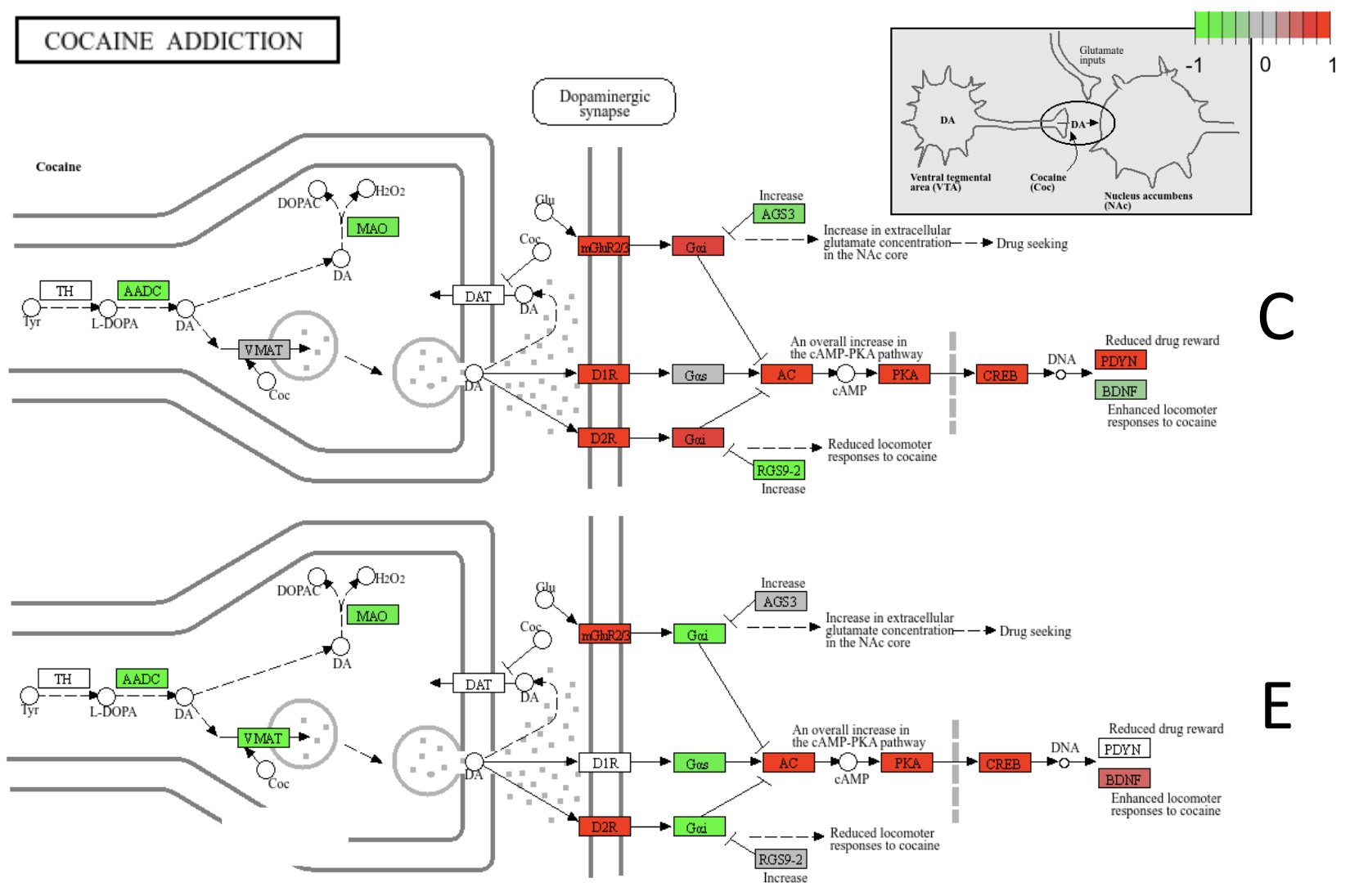
We defined the transcriptomic response of two compounds identified to inhibit HSF1 expression. The compounds display substantially different responses in gene expression, indicating their mechanisms of action are likely unique. Further analysis revealed a pattern of suppressed pathways involving metabolism/stress and activated involving apoptosis and dopaminergic paths. We predict that the compounds cause similar cellular responses through different mechanisms of action. The compounds also show similarities to D2R antagonists. Interestingly, D2R has been suggested to play a role in cancer proliferation, and several D2R antagonists are being considered for cancer drugs

Future Works
Future work will explore connections between HSF1, D2R, and cancer. The long-term goal is to explain how the compounds affect HSF1 to improve potency, explore side effects, and complete preclinical studies.


Genetic Investigation of Abnormal Ovulation in C. Elegans


Team Member(s)
Sara Sornat

Faculty Advisor
Dr. Karen S Kim Guisbert


Project Summary
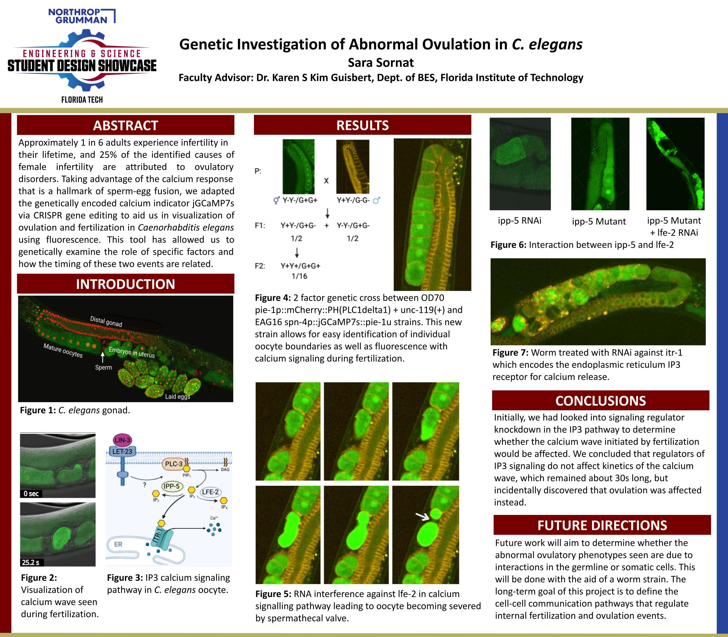
​​Approximately 1 in 6 adults experience infertility in their lifetime, and 25% of the identified causes of female infertility are attributed to ovulatory disorders. Taking advantage of the calcium response that is a hallmark of sperm-egg fusion, we adapted the genetically encoded calcium indicator jGCaMP7s via CRISPR gene editing to aid us in visualization of ovulation and fertilization in Caenorhabditis elegans using fluorescence. This tool has allowed us to genetically examine the role of specific factors and how the timing of these two events are related.


Analysis
Initially, we had looked into signaling regulator knockdown in the IP3 pathway to determine whether the calcium wave initiated by fertilization would be affected. We concluded that regulators of IP3 signaling do not affect kinetics of the calcium wave, which remained about 30s long, but incidentally discovered that ovulation was affected instead.

Future Works
Future work will aim to determine whether the abnormal ovulatory phenotypes seen are due to interactions in the germline or somatic cells. This will be done with the aid of a worm strain. The long-term goal of this project is to define the cell-cell communication pathways that regulate internal fertilization and ovulation events.


Isoform-specific effects of PYP-1 in Alzheimer's disease models and HSR regulation


Team Leader(s)
Rohan Gandhi Surapaneni

Team Member(s)
Rohan Gandhi Surapaneni

Faculty Advisor
Eric Guisbert


Isoform-specific effects of PYP-1 in Alzheimer's disease models and HSR regulation  File Download
Project Summary
The Heat Shock Response (HSR) is a universally conserved pathway that maintains cellular protein folding homeostasis, or proteostasis.  Tau protein misfolding and aggregation has a key role in the pathology of Alzheimer's disease, and HSR activation ameliorates Tau toxicity.  We have identified new genes that regulate the HSR using a genetic approach.  Among these genes is PYP-1, which is annotated as an inorganic pyrophosphatase and a component of the NuRF chromatin remodeling complex.  We have generated isoform-specific RNAi knockdown constructs for PYP-1 and tested the effects of these isoforms on HSR regulation and Tau toxicity.


Analysis
We made RNAi knockdown constructs of PYP-1 and its isoforms to study its effects on HSR tau toxicity, and egg-lay recovery after temperature stress.

Future Works
Validation of these proposed pathways using isoform overexpression. Investigation of other pathways/phenotypes for each isoform using RNA-seq. 


Biomedical Engineering

NMES Brace for Spasticity


Team Leader(s)
Emily Foley

Team Member(s)
Caitlin Streck, Kira Scarpignato, Emily Foley, Yasser Althobaiti

Faculty Advisor
Dr. Linxia Gu


Project Summary
The NMES Brace for Spasticity provides a non-invasive way to treat spastic posturing of the hand and wrist and improve patient mobility through dynamic bracing and electrical stimulation. Recent studies have shown greater improvement in patient mobility when dynamic bracing is coupled with targeted neuromuscular electrical stimulation [NMES]. NMES is the practice of providing electrical stimulation to muscle groups to elicit a contractile reaction. Dynamic bracing is the practice of not only providing structural support for an injury but also applying necessary corrective forces for optimal recovery. Despite its increased efficacy, there aren't any current products on the market that combine a dynamic brace with an electrical stimulation device into one easy, at-home therapy product. We propose an upper extremity dynamic brace that incorporates electrical stimulation in a safe, simple way that will be easy for patients to use daily in the comfort of their own homes.


Project Objective
The NMES Brace for Spasticity aims to tackle the challenges of current spasticity treatment options with a more patient-centric design, that enhances the comfort and ease of use at a lower cost than current commercially available treatments.

Manufacturing Design Methods
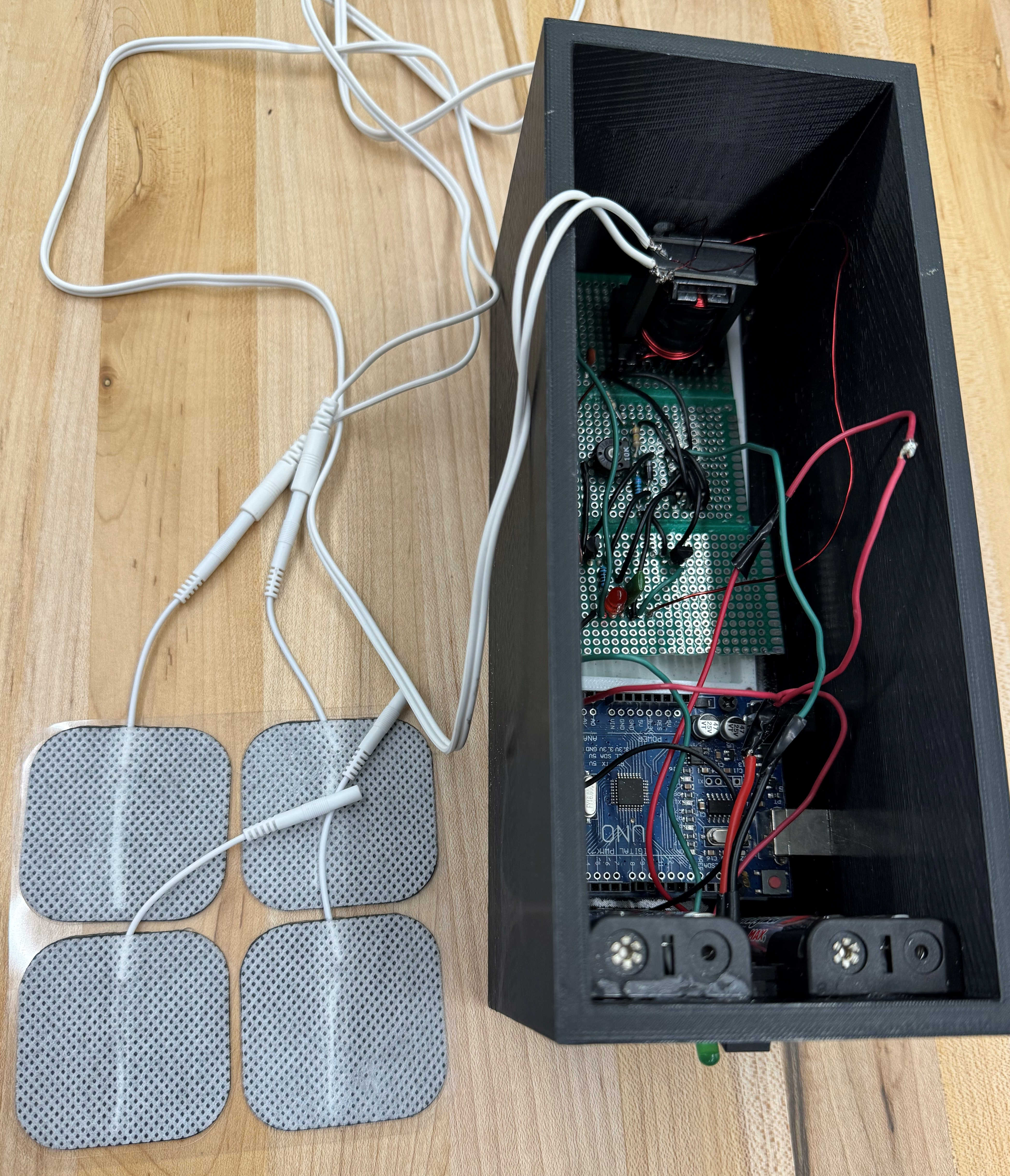
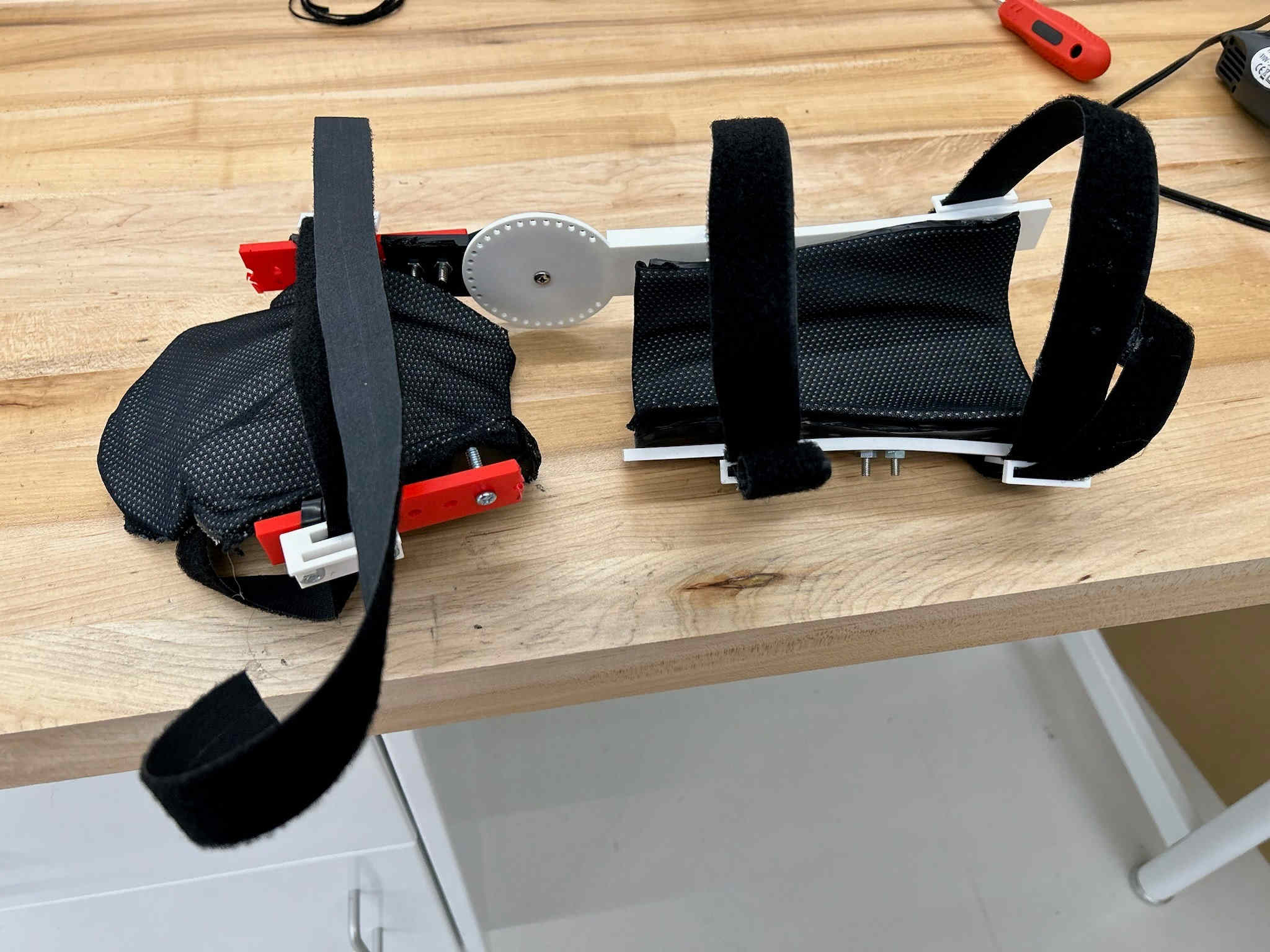
The brace is designed for the hand and wrist and uses thermoset plastic to mold a hand component and forearm component for the complete brace. The support bars and angle hinge were 3D printed. Then all parts were put together, with the support bars being screwed into the thermoset plastic pieces and velcro straps being added over the brace components. Thermoset molds were covered with felt and breathable mesh fabric for more comfort. The NMES circuit was constructed using an ICL7555 timer chip that provided a pulsatile output and custom-made transformer to get current outputs within a safe but sensible range for subject use. An Arduino Uno was integrated into the circuit and used to program an auto shut-off at the recommended time frame for safety purposes of 30 minutes.

Specification
The brace was designed so it could lock between 0 and 360 degrees, allowing for adjustment according to the needs of the patient. Output from the NMES circuit was a pulsatile frequency of 160 Hz, with a max amperage of 50 mA, a 50% duty cycle, and an auto shutoff after 30 minutes of stimulation.

Analysis
From the recorded data of subjects using the brace, data showed that it took less time for the subject to take off the brace only using one hand than putting it on. There was also no conclusive correlation between the brace comfort and the patient’s likelihood to continue the treatment method if they were a spasticity patient. However, subjects did remark that their likelihood to follow through on using the device was more based on their personal motivation than the design or possible effectiveness of the device. The NMES Brace for Spasticity is also cheaper than most commercially available treatment options.

Future Works
For future works on this project, the brace could be improved by changing the angle hinge design to metal for stability and durability while using the brace. Additionally, an orthopedic felt could be added to the thermoset plastic molds to add more comfort for the patients while they are wearing the device. The size of the NMES stimulation unit could be downscaled by printing the circuit components onto a PCB board. Following improvements of the device, physical therapists could be consulted for feedback on the design and evaluation of the likeliness of devices usage by their patients. Following an improvement to the design based on any feedback given, a patient trial would be conducted to verify the results of combined bracing and NMES to improve spasticity patient's range of motion and muscle strength.


Manufacturing Design Methods
The brace is designed for the hand and wrist and uses thermoset plastic to mold a hand component and forearm component for the complete brace. The support bars and angle hinge were 3D printed. Then all parts were put together, with the support bars being screwed into the thermoset plastic pieces and velcro straps being added over the brace components. Thermoset molds were covered with felt and breathable mesh fabric for more comfort. The NMES circuit was constructed using an ICL7555 timer chip that provided a pulsatile output and custom-made transformer to get current outputs within a safe but sensible range for subject use. An Arduino Uno was integrated into the circuit and used to program an auto shut-off at the recommended time frame for safety purposes of 30 minutes.


StrabiSense


Team Leader(s)
Karly Liebendorfer

Team Member(s)
Ingrid Cooper, Michael Ninomiya, Seda Yilmaz

Faculty Advisor
Dr. Linxia Gu


StrabiSense  File Download
Project Summary
Amblyopia, commonly known as lazy eye, affects approximately 4% of the U.S. population, translating to around 10 million individuals. This condition can develop over an individual's lifetime or may be present from birth, particularly in premature birth. Amblyopia is characterized by an imbalance in the muscle tension of the eyes, leading to misalignment and improper coordination between the two eyes. Traditional treatment involves strabismus surgery, which aims to correct this misalignment by adjusting the tension in the eye's muscles. While strabismus surgery primarily focuses on the misaligned eye, achieving symmetrical alignment and coordination between both eyes is the ultimate objective. However, adjustments to the misaligned eye muscles can sometimes inadvertently impact the unaffected eye. This interaction may lead to complications, including impaired vision in both eyes. To address these challenges, our team developed a force sensor, StrabiSense, explicitly designed for strabismus surgery. This innovative device will provide surgeons with real-time, quantifiable data on the force exerted by the extraocular muscles during the surgical procedure.


Project Objective
Our project aims to significantly advance the field of strabismus surgery by integrating a force sensor named StrabiSense into the standard surgical protocol. The primary objective is to provide surgeons with quantitative data to enhance the precision and effectiveness of surgical procedures. The StrabiSense is designed to measure the force exerted on the extraocular muscles during strabismus surgery and provide a quantitative assessment of muscle tension and deformation, enabling surgeons to make more informed decisions during eye muscle adjustment.

Manufacturing Design Methods
Our team redesigned the traditional strabismus surgery hook, introducing a compact, precision-enhanced device. This advanced tool features a shortened hook secured by a drill screw headpiece for optimal stability during procedures. A state-of-the-art strain gauge integrated beneath the hook mechanism is central to our design. This gauge accurately measures the force exerted on the eye muscle, transforming mechanical deformation into reliable data. As the hook engages the muscle, the device calculates the force applied. This information is displayed on an LED screen, guiding surgeons to tighten or loosen the muscle for optimal surgical outcomes. Our device represents a leap forward in surgical precision, offering real-time feedback that enhances patient safety and procedure efficacy. Our device is embedded with the Arduino Nano 33 BLE Sense, a module with a wireless connectivity feature. This enables integration with external devices, including computers and, shortly, a dedicated smartphone app. Our device facilitates real-time data transmission through this wireless capability, ensuring critical surgical information is always at the surgeon's fingertips.

Specification
Our surgical tool incorporates a modified strabismus surgery hook attached to a drill screw head bit. This component is precisely engineered to interact dynamically with our force sensor, facilitating the accurate force data exerted by the extraocular muscles during surgery. The force data is immediately transmitted to an Arduino Nano Sense BLE 33 microcontroller upon collection. The results of the data analysis are displayed via an LCD screen, which indicates the force levels through a color-coded system. A slide switch is incorporated into the design for power control, allowing surgeons to turn the device on or off as needed. The device includes a charging port on its base for enhanced convenience and usability in surgical environments.

Analysis
Our novel strabismus force sensor offers a groundbreaking approach to revolutionizing strabismus surgery. StrabiSense will streamline and standardize strabismus surgery by redefining the surgical landscape, ensuring accuracy and consistent results. The device is affordable without compromising quality or functionality. Our device is convenient, has wireless capabilities, and is portable since it does not require constant electrical outlet access, enhancing ease of use. Our sensor removes the guesswork from strabismus surgeries for training purposes, providing a reliable education and skill development tool. Our tool will allow strabismus surgeons to enhance surgical precision and a standardized procedure.

Future Works
Future improvements will emphasize refining the product's design to ensure it is more streamlined and visually appealing. The focus will be optimizing the device for ease of handling while integrating ergonomic principles to enhance user comfort and operational efficiency. Plans to develop an app designed explicitly for surgeon interaction. This advanced tool will facilitate real-time data exchange and decision-making support, enhancing surgical procedures' precision and effectiveness. Recognizing the paramount importance of sterilization in medical environments, our future models will incorporate state-of-the-art autoclavability features. Making the device smaller to cater to diverse medical scenarios. This development will further improve the ease of handling by medical professionals. It will also expand the device's applicability to a broader range of surgical environments and patient anatomies.


Manufacturing Design Methods
Our team redesigned the traditional strabismus surgery hook, introducing a compact, precision-enhanced device. This advanced tool features a shortened hook secured by a drill screw headpiece for optimal stability during procedures. A state-of-the-art strain gauge integrated beneath the hook mechanism is central to our design. This gauge accurately measures the force exerted on the eye muscle, transforming mechanical deformation into reliable data. As the hook engages the muscle, the device calculates the force applied. This information is displayed on an LED screen, guiding surgeons to tighten or loosen the muscle for optimal surgical outcomes. Our device represents a leap forward in surgical precision, offering real-time feedback that enhances patient safety and procedure efficacy. Our device is embedded with the Arduino Nano 33 BLE Sense, a module with a wireless connectivity feature. This enables integration with external devices, including computers and, shortly, a dedicated smartphone app. Our device facilitates real-time data transmission through this wireless capability, ensuring critical surgical information is always at the surgeon's fingertips.


Dance Dance Rehabilitation


Team Member(s)
Elizabeth Christ, Natalie Eustis, Griffin Martin, Collin Stewart

Faculty Advisor
Dr. Linxia Gu


Dance Dance Rehabilitation  File Download
Project Summary
The Dance Dance Rehabilitation project introduces a pioneering approach to neuromuscular rehabilitation in athletes, with a primary focus on concussion recovery. By developing an innovative concussion assessment tool that integrates interactive visual cues and precise neuromuscular measurements during dynamic activities like jumping, the project aims to address critical gaps in current concussion care protocols. The device itself is a sophisticated system comprising four force plates arranged in a configuration inspired by Dance Dance Revolution, accompanied by LED lights for visual cue stimuli. Each force plate consists of a top layer of clear acrylic, followed by a layer of plywood with load cells in each corner. These load cells, connected via a Wheatstone bridge to an Arduino Uno central control unit, accurately measure force production and time during jumping activities, from which response time can be deduced. This project holds significance as it not only enhances the understanding of neuromuscular dynamics post-concussion but also provides clinicians with a sophisticated platform to gauge athletes' readiness to return to play. By leveraging advanced technology and scientific inquiry, the project aims to usher in a new era of personalized rehabilitation strategies tailored to individual athletes' needs, ultimately contributing to improved athlete safety and well-being in sports-related activities.


Project Objective
The Dance Dance Rehabilitation project aims to develop a cutting-edge concussion assessment tool that integrates interactive visual cues and precise neuromuscular measurements during dynamic activities, with a primary focus on jumping. This system aims to accurately assess resposne times and force generation characteristics, providing clinicians with a user-friendly platform to gauge athletes' readiness to return to play post-concussion.

Manufacturing Design Methods
he device consists of four force plates arranged in a "T" configuration. Each plate features a clear acrylic top layer. Beneath the acrylic layer is a 3⁄4 inch plywood base with a laser-cut square to hold LED lights for stimulus cues. Load cells, serving as transducers, are positioned at each corner of the plywood base to measure force. These load cells are connected to an Arduino Uno, the central control unit, via an Hx-711 Amplifier equipped with a 24-bit analog-to-digital converter for precise data collection. The Arduino initiates stimuli through LED lights and collects force data from the load cells every 250 milliseconds. The collected data is then transferred to MATLAB for analysis, where force versus time graphs are generated to deduce peak force and response times for each movement.


Analysis
Data collected by the Dance Dance Rehabilitation device undergoes thorough analysis to derive key insights into athletes' neuromuscular function during concussion recovery. Utilizing MATLAB, load-versus-time graphs are generated to identify peak force and reaction times for each movement. These metrics are then averaged across the full test sequence of 10 movements for comprehensive evaluation, but each movement is also analyzed individually. A slow-motion video was taken of each test, and from that response times were deduced and compared to those from the Dance Dance Rehabilitation device for data validation.

Future Works
Future endeavors may focus on refining the device's hardware and software components to optimize data collection efficiency and accuracy. Additionally, expanding the device's capabilities to include a wider range of neuromuscular assessments and rehabilitation exercises could enhance its utility in clinical and athletic settings. Collaborative research efforts may also explore integrating machine learning algorithms to analyze data patterns and provide personalized rehabilitation recommendations. Continually, studies examining the device's long-term effectiveness in improving athletes' recovery outcomes could provide valuable insights into its clinical impact.


Manufacturing Design Methods
he device consists of four force plates arranged in a "T" configuration. Each plate features a clear acrylic top layer. Beneath the acrylic layer is a 3⁄4 inch plywood base with a laser-cut square to hold LED lights for stimulus cues. Load cells, serving as transducers, are positioned at each corner of the plywood base to measure force. These load cells are connected to an Arduino Uno, the central control unit, via an Hx-711 Amplifier equipped with a 24-bit analog-to-digital converter for precise data collection. The Arduino initiates stimuli through LED lights and collects force data from the load cells every 250 milliseconds. The collected data is then transferred to MATLAB for analysis, where force versus time graphs are generated to deduce peak force and response times for each movement.


3D SpectraTech


Team Leader(s)
Dyani Peterson

Team Member(s)
Danyelle Mohammed, John Panchookian, Dyani Peterson and Lexine Sibert-Jolissaint

Faculty Advisor
Dr. Linxia Gu


Project Summary
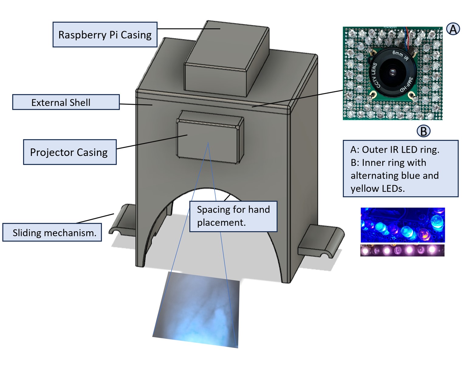
90% of patients in hospitals and clinical settings require intravenous (IV) cannulation to administer medicine or collect blood samples. However, the efficacy of IV cannulations can be hindered by various physical attributes, such as small vein diameter, deep vein location, dietary factors, medical history, venous disease, and skin pigmentation, which in turn makes vein detection challenging. Many devices were designed with this in mind to counteract these challenges; however, they do not allow for vein localization and depth, which oftentimes leads to medical practitioners losing sight of the veins and not knowing how deep they are. With these challenges in mind, 3D SpectraTech presents a spectroscopy-based vein detection system utilizing near-infrared (NIR) spectroscopy and LED sensor technology, designed to precisely image and locate veins within the arm and determine the corresponding depth through image processing, which then projects this image back onto the arm, enabling healthcare professionals to accurately identify veins, alleviating the uncertainty associated with vein localization. 3D SpectraTech stands out as a solution to these challenges by offering the capability to detect vein depth, distinguishing it from current market offerings and affordability, as current commercially available vein finders utilizing NIR technology are quite expensive, ranging from approximately $4500USD for portable versions to $27,000USD for non-portable equipment, rendering them inaccessible to many. The design of the device is broken into two components: the hardware and software systems. The hardware aspect consists of an external 3D-printed shell enclosing a camera surrounded by LED sensors. The LEDs are of three differing wavelengths, 450nm, 585nm, and 850nm, and are arranged in two layers. The hardware system captures the image of the vein, which then undergoes image processing in the software component. Image processing involves enhancing the image clarity, and once this is done, vein segmentation is done, which utilizes Python software programming to isolate veins to retrieve vein depth and thickness values. This segmented image is projected back onto the arm, where medical practitioners can now use the known vein depth value and the image projection to administer IVs or collect blood samples accurately.


Future Works
In future iterations of this project, several enhancements and developments can be made to improve further the functionality and usability of the vein detection system. Firstly, efforts will be directed towards miniaturizing the device and optimizing its design for mobility, allowing for easy transport and use in various medical settings, including remote or emergency situations. Additionally, minimizing the required distance between the camera and the target area will enhance the system’s efficiency and overall user experience. Lastly, the interface will provide feedback and adapt to different individuals with varying skin tones by implementing machine learning and an overall automated design. These future endeavors will elevate the vein-detecting system and ultimately advance its utility in clinical practice and patient care.


Innovative 4D Printing: Engineering Anisotropic Collagen-TCP Scaffolds for Bone Regeneration


Team Leader(s)
Karly Liebendorfer

Team Member(s)
Karly Liebendorfer

Faculty Advisor
Vipuil Kishore


Innovative 4D Printing: Engineering Anisotropic Collagen-TCP Scaffolds for Bone Regeneration  File Download
Project Summary
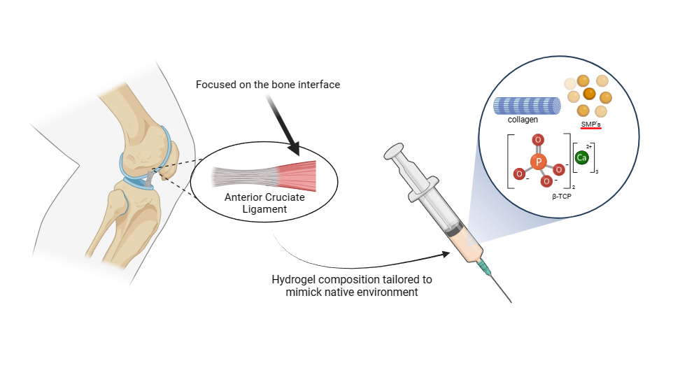
Composite biomimetic scaffolds fabricated by combining collagen with bioceramics show immense promise for bone tissue engineering (BTE) applications. In this realm, applications of extrusion-based 3D printing strategies with collagen-based composite inks yield 3D scaffolds with high precision. While these scaffolds could mimic the 3D complexity of native tissue, the microarchitectural aspects (i.e., collagen anisotropy) are lacking. Recent work in our lab has shown that combining extrusion-based 3D printing with magnetic alignment approaches in a 4D printing scheme triggers a high degree of collagen anisotropy in printed scaffolds. The current work aims to integrate a bioceramic and observe the effect on print fidelity, collagen alignment, and cellular responses.


Project Objective
This study aims to assess the impact of β-tricalcium phosphate (β-TCP) incorporation on the rheological properties of collagenous inks as well as on collagen anisotropy and print fidelity of 4D printed scaffolds. ​

Manufacturing Design Methods
The scaffolds were fabricated using methacrylated collagen (Col, 6 mg/ml) and xanthan gum (XG, 10 mg/ml) that were combined in a Col:XG ratio of 4:1, to which a cytocompatible photoinitiator (VA-086, 0.1% w/v), streptavidin-coated magnetic particles (SMP, 0.4 mg/ml), and β-TCP (0%, 10%, 70% w/w) were added. Collagenous constructs were 4D printed in the presence of a 0.2 T magnetic field and incubated for 30 min at 37 °C to induce gelation and crosslinked using UV light.

Specification
The print fidelity of the resultant scaffolds was measured using ImageJ by comparing the area of the printed scaffolds to that of the 3D model. Collagen fiber alignment in 4D printed scaffolds was assessed using polarized light microscopy (PLM) and scanning electron microscopy (SEM). AlamarBlue (AB) assay was performed to analyze the effect of collagen alignment on Saos-2 metabolic activity (N = 3/group/timepoint). Quantitative data were analyzed using one-way ANOVA with Tukey posthoc test. Statistical significance was set at p

Analysis
Higher β-TCP concentration led to a disruption in collagen alignment and cell viability. However, print fidelity was not significantly impacted by ceramic incorporation. Lastly, preliminary results demonstrate promise for using 4D printing methods to generate biomimetic scaffolds for BTE applications.

Future Works
The effects of β-TCP incorporation on osteogenic cell response are currently under investigation. Studies are currently underway to identify the optimal ink composition that will yield composite scaffolds with retention of collagen anisotropy.


Manufacturing Design Methods
The scaffolds were fabricated using methacrylated collagen (Col, 6 mg/ml) and xanthan gum (XG, 10 mg/ml) that were combined in a Col:XG ratio of 4:1, to which a cytocompatible photoinitiator (VA-086, 0.1% w/v), streptavidin-coated magnetic particles (SMP, 0.4 mg/ml), and β-TCP (0%, 10%, 70% w/w) were added. Collagenous constructs were 4D printed in the presence of a 0.2 T magnetic field and incubated for 30 min at 37 °C to induce gelation and crosslinked using UV light.