Mission

The mission of the Biomedical and Chemical Engineering and Sciences Department is to provide a safe working environment in the pursuit of excellence in education, research and innovation in the fields of biomedical science and engineering, chemistry and chemical engineering. 

Biological Sciences

Chlorophyll, carotenoid, and motility variation among Chlamydomonas strains under quorum-sensing conditions


Team Leader(s)
Marcel Pedrosa

Team Member(s)
Marcel Pedrosa


Secondary Faculty Advisor
Andrew George Palmer


Chlorophyll, carotenoid, and motility variation among Chlamydomonas strains under quorum-sensing conditions  File Download
Project Summary
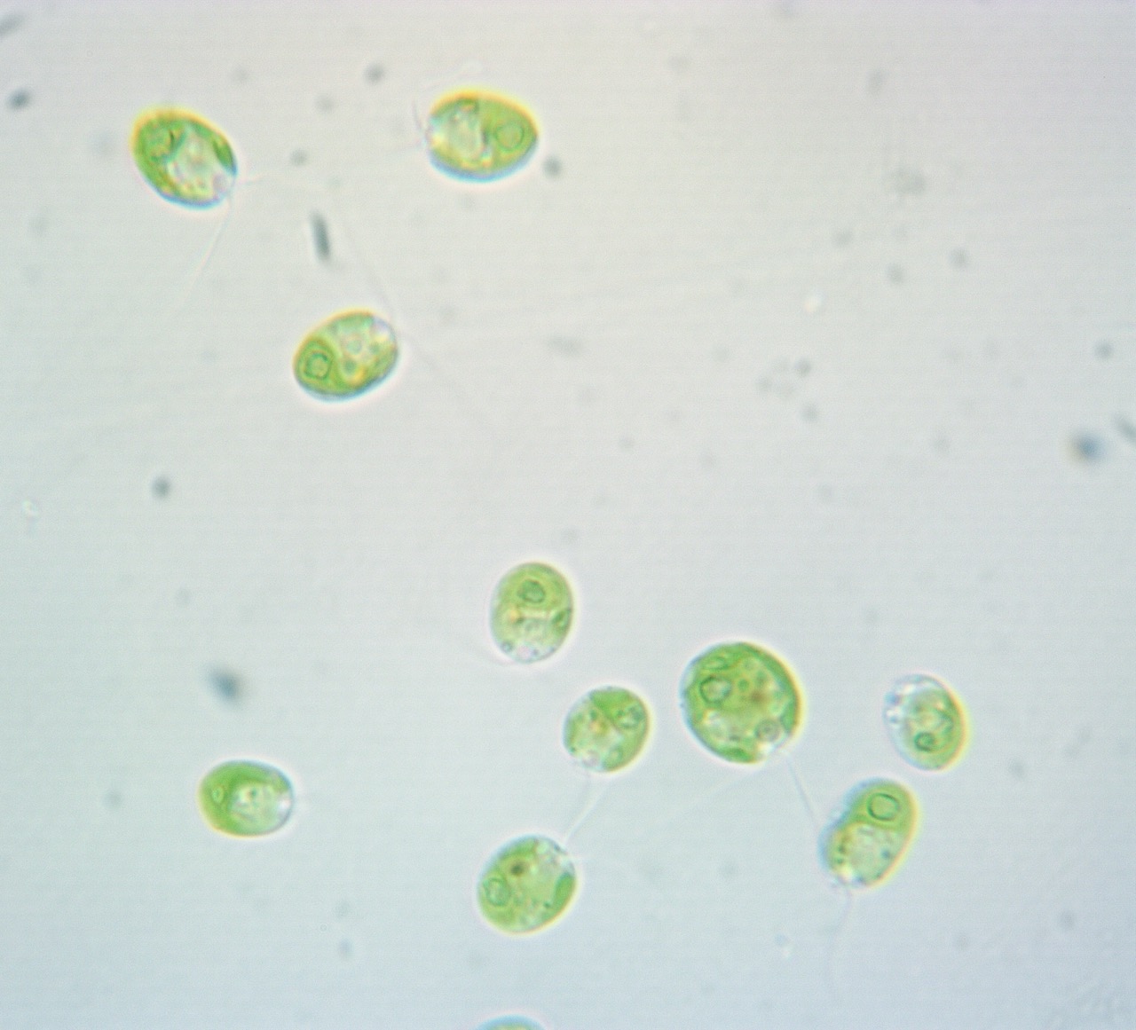
Quorum sensing (QS) is a cell-to-cell communication system that allows unicellular organisms to monitor their population density and synchronize behaviors using signaling molecules. Once a concentration threshold has been reached, the cells collectively alter/express gene expression, leading to a coordinated phenotypic switch, such as biofilm formation and virulence factor production. While predominantly found in prokaryotes, studies have confirmed this phenomenon is conserved among archaea and unicellular eukaryotes. This includes the model photosynthetic unicellular eukaryote Chlamydomonas reinhardtii, in which QS regulates swimming speed. This QS response shows some variation between ecological isolates of this species (strains), suggesting there may be differential responses between lines, as has been observed in some bacterial strains. This information indicates that Chlamydomonas reinhardtii could potentially utilize QS as a broader regulatory mechanism, coordinating changes in motility and metabolism in response to nutrient availability and population density. In this way, QS can facilitate cell distribution and adaptation towards environmental stress factors. With Chlamydomonas reinhardtii, they use photosynthetic pigments such as chlorophylls a and b and carotenoids to capture light for ATP and protect against oxidative stress, QS could help regulate their production as part of an adaptive response, while linking cell density and nutrient conditions to both motility and photosynthetic efficiency, which can lead to cultivation of optimized populations to survival under fluctuating environmental stress. Here, I will leverage an existing microplate assay to determine if there is a correlation between chlorophyll and carotenoid concentration with cell density, supporting a link to quorum sensing. Furthermore, an additional screen phenotypic screening will be conducted, in that model Chlamydomonas reinhardtii will be grown in different media with varying amounts of nitrogen then measured for cell density as well as chlorophyll and carotenoid content. These findings will assist in furthering our understanding of quorum sensing, showing in particular what traits it may influence with Chlamydomonas reinhardtii, specifically if it is with the production of chlorophyll and carotenoids, and if its trigger can be tied to specific environmental factors, such as low or high nutrient availability.


Project Objective
The goal of this study is to evaluate whether QS in Chlamydomonas reinhardtii is regulated by nutrient availability. From testing this, we can either support or refute this claim, thereby uncovering potential applications in biotechnology—such as optimizing algal production for biofuel or pigment synthesis—while providing deeper insight into microbial communication and survival strategies in nutrient-limited environments.

Manufacturing Design Methods
Six cultures were prepared in total, with three controls and three experimental replicates. All six cultures were grown in sterile 250 mL flasks containing 50 mL of either TAP media (control flasks) or nitrogen-depleted (N-TAP) media (replicate flasks). A single colony of strain cc124 is aseptically transferred into each flask under sterile conditions in a fume hood to prevent contamination. Both the control and replicant flasks were then monitored over 144 hours at set intervals (every 24 hours), during which multiple parameters were measured. During this period, parameters such as cell growth rate (cell density over each 24-hour interval), Pigment concentrations (chlorophyll A, chlorophyll B, and carotenoids per cell over each 24-hour interval), and the swim speed of the cell population were measured at an LCD period (at hour 24) and an HCD period (at hour 120).

Specification
To prepare TAP media suitable for Chlamydomonas reinhardtii, for 1-liter preparation, the media must for 1-liter preparation, the media must consists of 20 mL of 1 M Tris base (prepared by dissolving 30.14 g Trizma base in 250 mL DI water), 1.0 mL of phosphate buffer II (made from 10.8 g K₂HPO₄ and 5.6 g KH₂PO₄ in 100 mL), 10.0 mL of Solution A (containing 20 g NH₄Cl, 5 g MgSO₄·7H₂O, and 2.5 g CaCl₂·2H₂O in 500 mL), 1.1 mL of Hutner’s trace elements, and 1.0 mL of glacial acetic acid, with the remaining volume brought to 1 L using DI water. The final pH is adjusted to approximately 7.0 to support optimal algal growth. To prepare nitrogen-reduced TAP (N-TAP) media suitable for Chlamydomonas reinhardtii, for 1-liter preparation, the media must consists of 20 mL of 1 M Tris base (prepared by dissolving 30.14 g Trizma base in 250 mL DI water), 1.0 mL of phosphate buffer II (made from 10.8 g K₂HPO₄ and 5.6 g KH₂PO₄ in 100 mL), 10.0 mL of modified Solution A (containing 5 g NH₄Cl, 5 g MgSO₄·7H₂O, and 2.5 g CaCl₂·2H₂O in 500 mL), 1.1 mL of Hutner’s trace elements, and 1.0 mL of glacial acetic acid, with the remaining volume brought to 1 L using DI water. The final pH is adjusted to approximately 7.0 to support optimal algal growth.

Analysis
The growth rate (cell density) and Pigment concentrations (chlorophyll A, chlorophyll B, and carotenoid) per cell were quantified using a spectrometer, where samples were loaded into well plates and absorbance readings were taken at specific wavelengths (470 nm, 550 nm, 647 nm, and 663 nm). Blank readings from media alone were also collected to correct for background absorbance. The swim speed of the cell population was assessed by recording videos of live cells under a microscope at an LCD period (hour 24), then an HCD period (hour 120), and later analyzing them using the Fiji program (R-based tracking software). All collected data from the trials were organized in spreadsheets and analyzed using R code to calculate averages and interpret trends across conditions.

Future Works
Further studies into Chlamydomonas reinhardtii, will examine how its QS response to environment conditions, is affected by interchanging day-night cycle as they do have a natural circadian rhythm, which play a role in gene regulation.


Acknowledgement
Haire, T. C., Bell, C., Cutshaw, K., Swiger, B., Winkelmann, K., & Palmer, A. G. (2018). Robust microplate-based methods for culturing and in vivo phenotypic screening of Chlamydomonas reinhardtii. Frontiers in Plant Science, 9. https://doi.org/10.3389/fpls.2018.00235 Lee, D. Y., Park, J.-J., Barupal, D. K., & Fiehn, O. (2012). System response of metabolic networks in Chlamydomonas reinhardtii to total available ammonium. Molecular & Cellular Proteomics, 11(10), 973–988. https://doi.org/10.1074/mcp.m111.016733 Virtanen, O., Khorobrykh, S., & Tyystjärvi, E. (2020). Acclimation of chlamydomonas reinhardtii to extremely strong light. Photosynthesis Research, 147(1), 91–106. https://doi.org/10.1007/s11120-020-00802-2 Figure 1 created in https://BioRender.com


Multi-Strain Assessment of Quorum Sensing in Chlamydomonas reinhardtii


Team Leader(s)
Jaqueline Krueger

Team Member(s)
Jaqueline Krueger

Faculty Advisor
Dr. Andrew Palmer


Multi-Strain Assessment of Quorum Sensing in Chlamydomonas reinhardtii  File Download
Project Summary
Quorum sensing (QS) allows microbial populations to coordinate phenotypic switching, selecting for specific behaviors that are only beneficial when expressed at high cell densities (HCD, 106 cells/mL) rather than low cell densities (LCD,


Project Objective
To investigate the conservation of quorum sensing across sequenced strains of C. reinhardtii. It is hypothesized that quorum sensing will vary across different strains of C. reinhardtii. These variations can then be leveraged to identify potential candidates for the identity of the QSMs as well as the relevant synthase(s) and receptor(s).

Manufacturing Design Methods
The swim speeds videos for high and low cell densities cultures of C. reinhardtii were collected via a light microscope equipped with a camera. In addition, their cell densities were verified via a hemocytometer. The cell cultures were placed into a centrifuge to separate the cells from the media. The media and the cells were then swapped with those of the opposite density and left to incubate for about 3 hours. Afterwards, videos were again taken using the same procedure as the controls. Specifications: Strains examined included CC-124, CC-408, CC-1690, CC-2343, CC-3269, and CC-4414.

Specification
Strains examined included CC-124, CC-408, CC-1690, CC-2343, CC-3269, and CC-4414.

Analysis
Initial video analysis was done using the TrackMate plugin for ImageJ. The swim speeds of the cells data are extracted from the videos and used to create violin plots and box plots. These plots are used for visual analysis of the data. If the procedure resulted in an increase in swim speed in a given strain following the media swap it indicates that the strain employs quorum sensing to mediate swim speed.

Future Works
The diversity of quorum sensing responses across strains of C. reinhardtii may be leveraged to characterize the signal and molecular elements of this phenomenon in this cosmopolitan species. Further research will investigate the mechanisms that dictate swim speed in CC-1690. On-going genetic analysis of CC-4414 may identify a receptor for this signal.


Acknowledgement
I would like to thank Dr. Palmer for his considerable help with this project. He offered invaluable advice and feedback throughout my research. I am forever grateful for the opportunities he has given me. I would also like to thank Ryan Quick and Lucy Turner helping for laying the ground work of this project and teaching me the ropes. Additional thanks to Ryan for assisting me when I needed it most during the long hours of experiments.


Biomedical Engineering

An Engineered Platform For Detecting Cellular Damage Upon Mechanical Impact


Team Leader(s)
Mai Tran

Team Member(s)
Lindsay Avila, Nina Fiordaliso, Mai Tran

Faculty Advisor
Dr. Linxia Gu

Secondary Faculty Advisor
Dr. Pengfei Dong


Project Summary
Mechanical forces play an important role in influencing cellular behavior, particularly through mechanotransduction, where physical stimuli are converted into biological signals. These forces can alter cellular stress responses, protein expression, and overall cell viability. While previous studies have examined factors such as strain, pressure, and acceleration, there remains a need for simplified in vitro systems that directly link controlled mechanical impact to measurable cellular responses. In this study, we developed an experimental platform to apply controlled mechanical impacts to cultured cells in a small-scale environment. Cells were subjected to varying levels of impact, including control, moderate, and high-impact conditions. Heat shock protein 70 (HSP70), a biomarker associated with cellular stress, was measured using an ELISA assay, and cell viability was assessed using a live/dead assay. Results showed that mechanical impact significantly influenced HSP70 expression, with moderate impact conditions producing a measurable decrease compared to control. However, the highest impact condition exhibited increased variability and did not follow a consistent trend. In contrast to the initial hypothesis, live cell counts increased across all impact conditions, indicating that the applied mechanical forces did not induce widespread cell death. These findings suggest that mechanical impact alters cellular stress responses without necessarily compromising viability, and that the applied conditions were likely sub-lethal. This study shows how controlled mechanical forces influence cellular behavior and offers a platform for investigating stress-response mechanisms in vitro.


Project Objective
Design an in vitro platform to quantitatively assess the effects of progressively increasing controlled mechanical impacts. Evaluate cellular stress response through the HSP70 expression and cell viability using a live/dead assay. Investigate the relationship between mechanical impact, cellular stress response, and cell viability

Manufacturing Design Methods
Impact: Seed NIH-3T3 cells in appropriate culture plates and incubate under standard conditions (37 °C, 5% CO₂) such that cell confluency reaches approximately 70–80% on the day of the experiment. On the experiment day, subject each sample group to mechanical impact. For a single impact trial, maintain a distance of 2 cm between the bottom of the 35 mm petri dish and the impact source. For multiple impact trials (2-impact and 3-impact conditions), repeat the impacts immediately one after another without delay. Immediately after impact, replace the culture media with non-FBS media. Return the cells to the incubator and allow them to recover for 24 hours. After the 24-hour incubation period, collect the conditioned media from each sample for ELISA analysis. ELISA Procedure: Set standard, test sample, and control (zero) wells on the pre-coated plate and record their positions. It is recommended to measure each standard and sample in duplicate. Wash the plate 2 times before adding reagents. Aliquot 0.1 mL of standard solutions into the standard wells. Add 0.1 mL of Sample/Standard dilution buffer into the control (zero) well. Add 0.1 mL of collected samples into test sample wells. Seal the plate and incubate at 37 °C for 90 minutes. Remove the cover and discard the plate contents. Tap the plate on absorbent material. Do not allow wells to dry. Wash the plate 2 times. Add 0.1 mL of Biotin-detection antibody working solution into each well. Add to the bottom without touching the side walls. Seal and incubate at 37 °C for 60 minutes. Remove the cover and wash the plate 3 times with wash buffer, allowing buffer to sit for 1 minute each time. Add 0.1 mL of SABC working solution into each well. Cover and incubate at 37 °C for 30 minutes. Remove the cover and wash the plate 5 times with wash buffer, letting buffer remain in wells for 1–2 minutes each wash. Add 90 µL of TMB substrate into each well. Incubate at 37 °C in the dark for 10–20 minutes. Color development (blue) should be visible in higher concentration standards first. Add 50 µL of Stop solution to each well and mix thoroughly. The color will change to yellow immediately. Measure optical density (O.D.) at 450 nm using a microplate reader immediately after adding the stop solution. Live/Dead Assay: Following ELISA, perform a live/dead cell viability assay on the remaining cells. Prepare staining solution by adding 5 µL Calcein AM (Component A) and 20 µL Ethidium homodimer-1 (Component B) to 10 mL DPBS. Remove the culture medium from the cells. Add 100–200 µL of the staining solution directly to the cells. Incubate for 30 minutes at 20–25 °C. Measure fluorescence signals using a plate reader: Calcein AM: Excitation 494 nm / Emission 517 nm Ethidium homodimer-1: Excitation 528 nm / Emission 617 nm


Analysis
HSP70 concentrations were measured across control and increasing mechanical impact conditions using ELISA. A one-way ANOVA revealed a significant effect of impact condition on HSP70 levels (F(3,28) = 3.03, p = 0.046). Post hoc Welch’s t-tests comparing each impact group to control showed that Impact 1 (p = 0.028) and Impact 2 (p = 0.048) resulted in significantly lower HSP70 concentrations relative to control, while Impact 3 was not significantly different (p = 0.45). Mean HSP70 levels were highest in the control group and decreased in Impact 1 and Impact 2 conditions, with Impact 3 showing increased variability and a partial recovery in HSP70 levels. Cell viability was assessed using a live–dead assay, where live cell counts were quantified across the same conditions. Contrary to the initial hypothesis, live cell counts increased in all impact groups compared to control, with the highest counts observed in Impact 2. These findings indicate that while mechanical impact significantly alters HSP70 expression, it does not correspond to decreased cell viability under the tested conditions. Instead, the results suggest that the applied mechanical impacts were sub-lethal and may have stimulated cellular activity or survival responses rather than inducing widespread cell death.

Future Works
Deliver impacts to multiple contact points on cultured cells to assess stress response. Determine the threshold of sub-lethal vs. lethal impact on cells. Post-training vibration stimulation to promote muscle recovery. Acknowledgement: Dr. Linxia Gu, Dr. Pengfei Dong, Dr. Robert Usselman, Dr. Mohammad Ahmed, Dr. Christopher Bashur, Jianing Wang, Beste Caner.


Biopulse


Team Leader(s)
Andrew Carr

Team Member(s)
Andrew Carr, Stefan Paton, Jan Deniz, Rachel Hizon

Faculty Advisor
Linxia Gu


Project Summary
A Split Hopkinson (Kolsky) Bar (SPHBs) is a device used to find a material’s mechanical response under high strain rate loading scenarios (10² – 10⁴ s⁻¹). It is commonly used to simulate vehicle crashes or ballistic impacts. The novelty of the device is its size and cost of its components compared to other SPHBs on the market. The motivation is to further the capabilities on campus by providing the first dynamic material testing device.


C-SIDE


Team Leader(s)
Emily Acampora

Team Member(s)
Emily Acampora, Emma Angelo, Kyle Burden

Faculty Advisor
Dr. Linxia Gu

Secondary Faculty Advisor
Dr. Careesa Liu


C-SIDE  File Download
Project Summary
This project focuses on developing a rapid concussion diagnostic system using objective physiological measures recorded with the Muse 2 wearable headset. Current concussion assessments rely heavily on subjective symptom reporting and time-intensive evaluations, which can lead to underdiagnosis and unsafe return-to-play decisions. To address this gap, our system leverages measurable physiological signals, recording brainwave activity, heart rate, and head movement data to provide a more accurate and reliable assessment of brain function. Participants complete a series of short tasks to assess the cognitive deficits, autonomic dysregulation, and postural instability known to be associated with concussions. The data is streamed automatically through a user interface, where it automatically processes and extracts relevant biomarkers. The overall goal is to create a fast, non-invasive, and user-friendly diagnostic tool that can be deployed in real-world athletic settings. Ultimately, this approach aims to improve diagnostic accuracy, support safer return-to-play decisions, and reduce the long-term risks associated with undiagnosed or repeated concussions.


Project Objective
Develop a fast, objective method for concussion detection using physiological data, integrating EEG, PPG, and motion data into a single, synchronized assessment system. Design a quick and user-friendly testing protocol for real-time use. Reduce reliance on subjective symptom reporting. Improve diagnostic accuracy and support safer return-to-play decisions.

Manufacturing Design Methods
Athletes from contact and non-contact sports at Florida Tech were recruited. Each participant completed a baseline assessment, with additional testing for those who sustained a concussion. Data was collected using the Muse 2 headset. Participants completed three short tasks (Go/No-Go, 2-back, postural balance), lasting a total of 5.5 minutes. All data was streamed in real time to a custom platform using Lab Streaming Layer (LSL). Data were filtered, segmented, and analyzed, extracting relevant features for comparison. Data were stored through a secure, cloud-based platform with user-specific access for athletes and trainers. The system integrates real-time physiological data with baseline comparisons to support fast, objective concussion assessment.


Future Works
Future efforts will focus on expanding the dataset to include a larger and more diverse population of athletes, particularly individuals who have sustained concussions. Increasing the sample size will strengthen the reliability and generalizability of the results. Additionally, a more robust dataset will support the development of machine learning models capable of identifying concussion-related patterns through features extracted from EEG, PPG, and gyroscope data, further enhancing the accuracy and scalability of the diagnostic system.


Acknowledgement
Dr. Careesa C. Liu, Dr. Mohammad Ahmed, Dr. Linxia Gu, Florida Tech Athletics


Cardio Clarity


Team Leader(s)
Emily Matheson

Team Member(s)
Emily Matheson, Matthew Kotze, Caroline Moore, and Kaitlyn Dunn

Faculty Advisor
Dr. Linxia Gu


Cardio Clarity  File Download
Project Summary
Cardio Clarity is a wearable device that monitors the heart’s electrical (electrocardiogram) and mechanical (seismocardiogram) activity, providing a more complete picture of cardiac health. Early monitoring has been shown to prevent up to 80% of cardiac events. This project focuses on designing a cost‑effective, multimodal system that can detect relevant cardiac features and present them in an accessible, real‑time interface.


CardioSync


Team Member(s)
Brendan Bouchard, Morgan Brenner, Panagiotis Vasileiadis, Michaela Wittman


CardioSync  File Download


OcuSensus


Team Member(s)
Asa Barnhart, Niko Burger, Patricia Carrelo-Creque, Kristie Fonseca

Faculty Advisor
Dr. Linxia Gu

Secondary Faculty Advisor
Dr. Pengfei Dong


Project Summary
Strabismus, commonly referred to as "lazy eye", is a condition characterized by the misalignment of the eyes, and affects approximately 2-5% of the general population. This condition can be treated through (strabismus) surgery, in which a surgeon carefully readjusts the patient's extraocular muscles into the correct position. However, this procedure is not a long-term solution for many, with about 36% of patients requiring reoperation within 5 years of the initial surgery. This is likely the result of insufficient force application on the extraocular muscles during surgery, as current methods rely solely on the intuition and skill of the performing surgeon. OcuSensus is a device created to combat this issue by providing real time qualitative feedback to performing surgeons. The device is equipped with red, yellow, and green LEDs that let the surgeon know if they are applying an appropriate amount of force. The goal for this qualitative feedback system is to allow the surgeons to fully correct a patient's strabismus with a higher retention rate than with the typical method of intuition-based correction.


VitaSense


Team Leader(s)
Ava Dorow

Team Member(s)
Ava Dorow, Ashley Schindler, Justin McParland

Faculty Advisor
Dr. Linxia Gu

Secondary Faculty Advisor
Dr. Sujoy Ghosh Hajra


VitaSense  File Download
Project Summary
An imaging photoplethysmography (iPPG) pipeline is a fully automated MATLAB-based system used to extract physiological vitals like heart rate (HR), blood oxygen saturation (SpO2), and blood pressure from standard video recordings of a subject's face without any physical contact. It operates by detecting subtle, periodic color variations in facial skin caused by underlying blood flow, processing those signals through multiple extraction algorithms, spectral analysis, and filtering to produce accurate vital sign estimates. The system's motivation is to enable low-cost, non-invasive vital sign monitoring using only a camera, with validation against clinical-grade ECG ground-truth hardware.