


Equipment
Equipment used by the Biomedical Engineering and Science department in its teaching and research.
Compiled by Lab Manager Beste Caner.
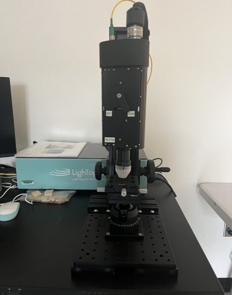
Uses a combination of Optical Coherence Tomography (OCT) and Gabor Domain Optical Coherence Microscopy (GDOCM). OCT can provide cross-sectional images of tissue structure on the micron scale in situ and in real time.
Phase contrast is a light microscopy technique used to enhance the contrast of images of transparent and colorless specimens. It enables visualisation of cells and cell components that would be difficult to see using an ordinary light microscope.
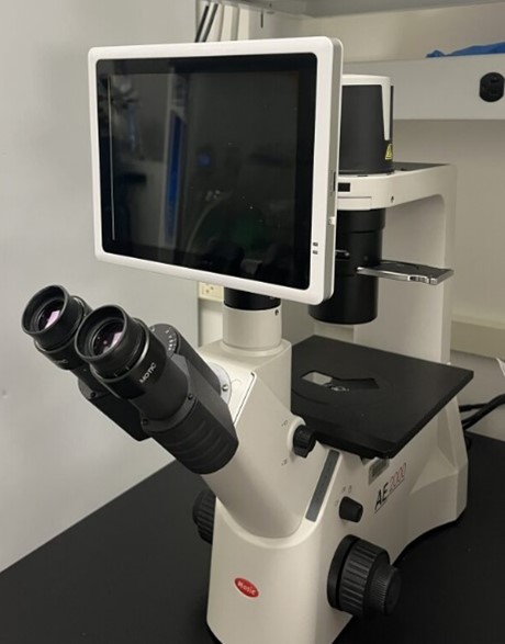
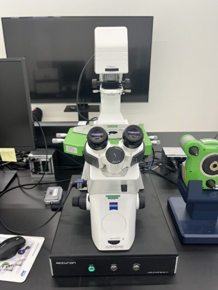
Motic's new AE2000 Inverted Microscope is designed for routine live cell inspection. In order to see the cells inside of the petri dish or tissue culture flask, inverted microscope is used where the objective is underneath the stage of the microscope and points upwards so that the specimen can be viewed from below.

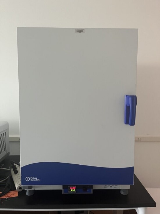
Provides accurate heating for routine laboratory procedures, drying and staining of slides, paraffin embedding, tissue culture work, incubation of antibody tests, microbiological determinations, crystallization studies and more.
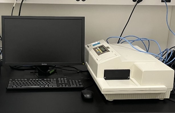
A microplate reader is a laboratory instrument that is used to measure chemical, biological or physical reactions, properties, and analytes within the well of a microplate.
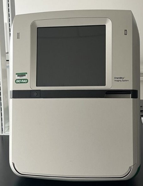
The ChemiDoc MP Imaging System is an instrument for imaging and analyzing gels and western blots. It is designed to address multiplex fluorescent western blotting, chemiluminescence detection, general gel documentation applications, and stain-free technology imaging needs.
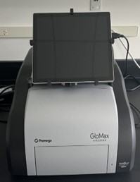
An all-in-one fluorescence imaging microscope that captures clear images with full electronic control and no darkroom required. This system allows live cell imaging, high-resolution observation of entire tissues.

Cell sorting involves physically separating (purifying) a specific cell population from the remaining cells or particles in the suspension for the purpose of performing further experimental procedures on these purified cells. The system is ideal for sorting cells expressing fluorescent proteins such as eGFP, DsRed, and mCherry, or for cells labeled with fluorescent markers such as antibodies labeled with PE, PE tandems, and other fluorophores excited by the 405, 488, 561, and 640 nm lasers.
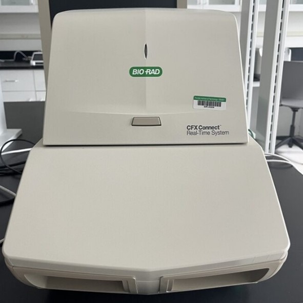
Real time PCR can be used for different applications such as gene expression analysis, microRNA & Noncoding RNA Analysis, genetic variation analysis, mutation detection, etc.
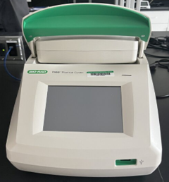
The thermal cycler (also known as a thermocycler, PCR machine or DNA amplifier) is a laboratory apparatus commonly used to amplify segments of DNA via the polymerase chain reaction (PCR).
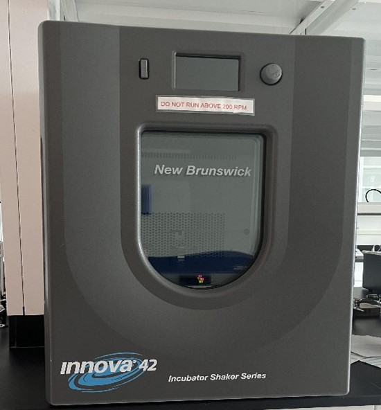
Shaking incubators are often used for cell culturing, cell aeration, and solubility studies. Eliminating the need of placing a separate shaker inside an incubator, the instruments incorporate oxygen and evenly distribute nutrients throughout the culture media.
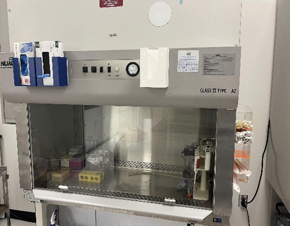
Biological Safety Cabinets (BSCs) are among the most common and effective primary containment devices used in laboratories to protect personnel against biohazardous or infectious agents and to help maintain quality control of the material being worked with as it filters both the inflow and exhaust air.
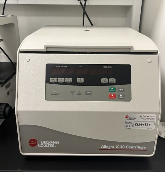
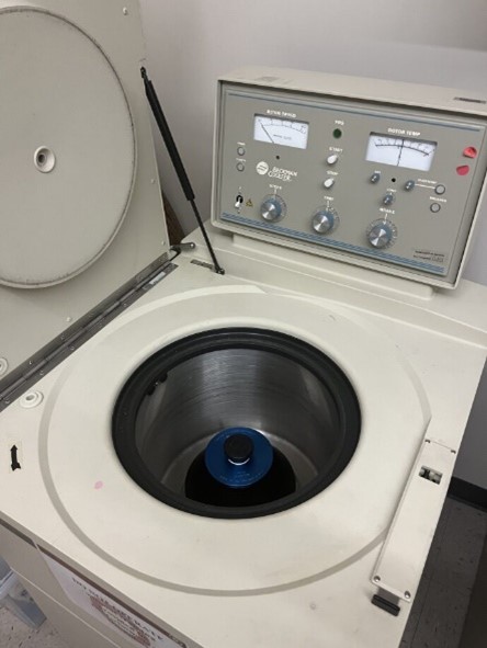
Centrifuges separate or concentrate substances suspended in a liquid medium by density and allows cell culture processing, blood sample preparation, and microplate applications.
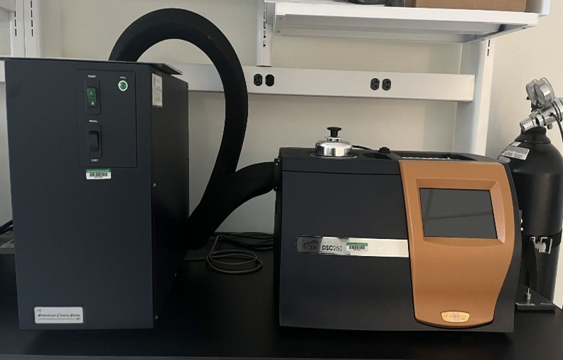
Measures how physical properties of a sample change, along with temperature against time. In other words, the device is a thermal analysis instrument that determines the temperature and heat flow associated with material transitions as a function of time and temperature.
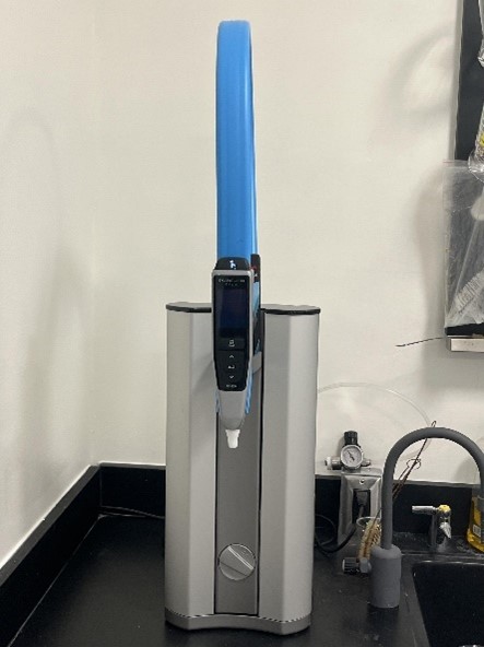
The flex 3 is the ultimate system providing tap to ultrapure water in one single unit and designed for a range of applications including electrochemistry, atomic spectroscopy, IC (Ion Chromatography), liquid chromatography, immunochemistry, HPLC , molecular biology, plant tissue culture, electrophoresis & mammalian cell culture.
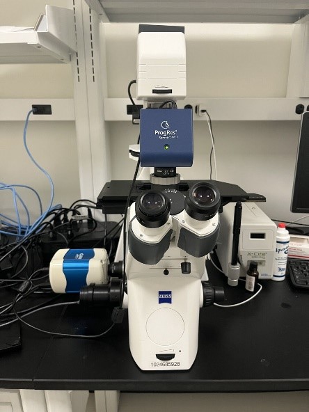
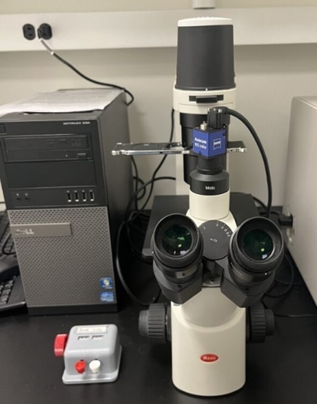
Jenoptik and Photometrics CoolSNAP MYO camera; and with Image-Pro (version 7.0) software. An inverted fluorescent microscope uses fluorescence to study specimens, such as living cells at the bottom of a petri dish or tissue culture. Fluorescence microscopy has ability to use fluorescence when the samples are labeled/stained with a fluorescent substance such as a dye antibody or protein allowing images to have contrast. By targeting these fluorescent labels, researchers can select what they want to see.
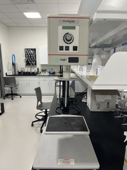
Homogenizers function as mixers that reduce particle size or force immiscible liquids to mix.
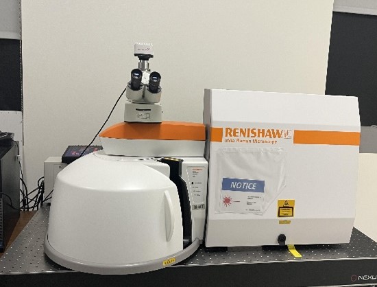
Raman spectroscopy is a powerful tool for determining chemical species. As with other spectroscopic techniques, Raman spectroscopy detects certain interactions of light with matter. Raman microscopy can be used for chemical or molecular analysis of unknown compounds in a small area, down to less than a micron.
Multi-mode microplate readers are used in different research fields for the quantification of different biological and chemical assays in a microplate. Typical applications include ELISA, nucleic acid, protein, enzymatic type homogeneous and heterogeneous assays, microbial growth, endotoxin testing, and pipettor calibration.
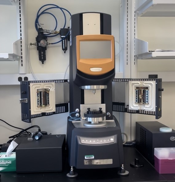
Rheology testing is measuring the deformation of matter under the influence of imposed stress, by analyzing the internal response of materials to forces. The temperature control design provides fast response and temperature stability over a continuous range of 760°C
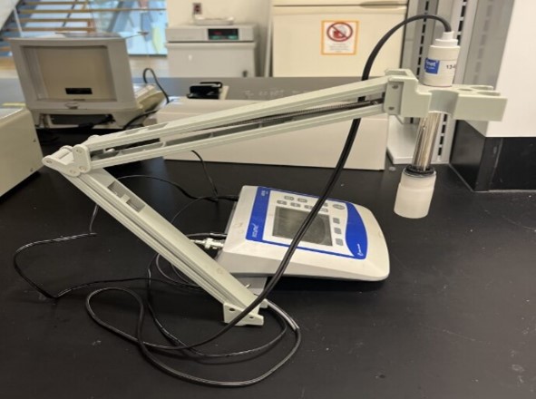
A benchtop pH meter is an electronic instrument used to measure the acidity, alkalinity of liquid, conductivity, ion concentration, ORP, and dissolved oxygen.
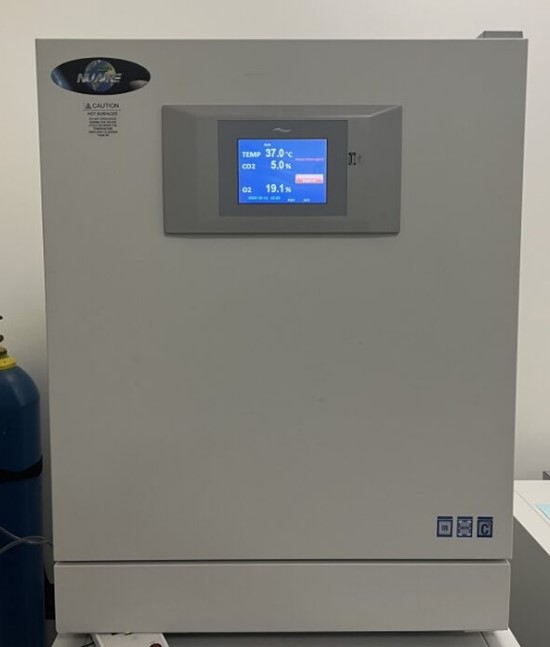
Cell culture incubator provides a reliable in-vitro environment for optimum cell culture growth, as well as the storage and preservation of embryos, gametes, and animal tissue cell cultures.
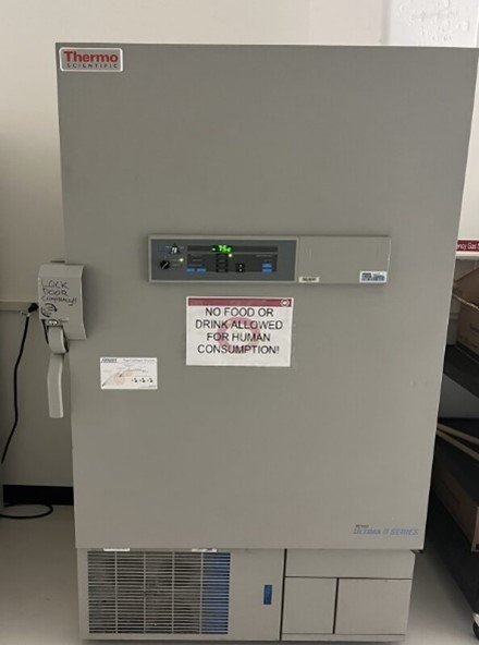
Developed for the most critical material storage. Store reagents, pharmaceuticals, biologicals and other commonly used laboratory materials.
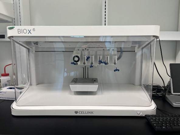
The BIO X bioprinter ensure the combination of 3D printing with biomaterials to replicate parts that imitate natural tissues, bones, and blood vessels in the body. Bioprinters print with cells and biomaterials, creating organ-like structures that let living cells multiply.
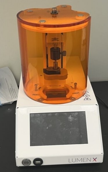
The Lumen X leverages digital light processing (DLP) printing to offer users high resolution, high throughput and high fidelity, enhancing applications in microfluidics, cell-laden hydrogels, macroporous structures and more.
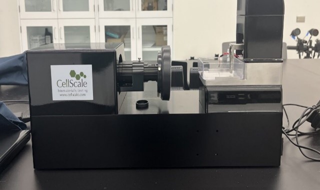
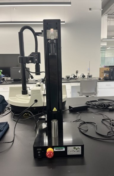
This system allows to do compression, tension, bending, indentation and shear testing with better force resolution. Applications include small tissue samples, hydrogel microspheres, cell spheroids, and engineered microtissues.
.jpg)
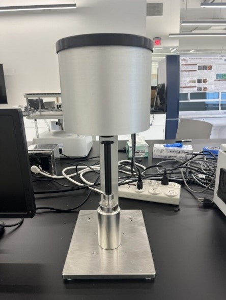
Biaxial testing is critical for understanding the mechanical properties of biomaterials due to their directionally oriented microstructures.
The system is capable of tension, compression, and bending testing using a wide range of grips and fixtures to accommodate different specimens and testing modes.
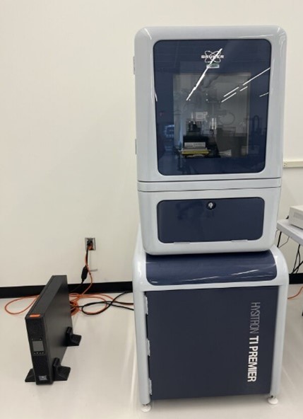
Enables nano-to-micro indentation, nanoscratch, nanowear, dynamic nanoindentation, in-situ SPM imaging and high-speed property mapping. Provides the broadest range of innovative characterization techniques, universal sample mounting options and modular system architecture.
The AFM can be used to image the topography of soft biological materials in their native environments.
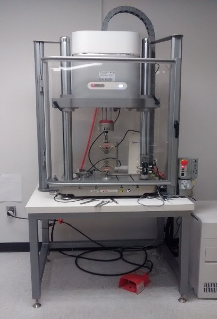
Compression machine is used to measure the compressive strength of a material. This is designed to apply a compressive load to the sample until it fails.
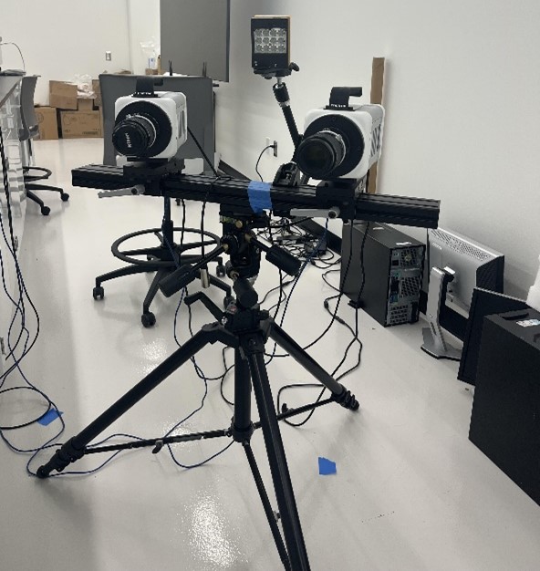
A high-speed camera is a device capable of capturing moving images with exposures of less than 1/1,000 second or frame rates in excess of 250 fps. It is used for recording fast-moving objects as photographic images onto a storage medium.
Instron ElectroPulse E3000 – mechanical testing system, using 3 special software: Instron Console, WaveMatrix and Bluehill. The Electropulse offers both linear and rotary motion which can be applied through a wave function with a variety of shapes and a range of frequencies so it can be used for precise fatigue testing of tensile/compressive loads, torsional loads, and 3-point bending.
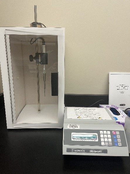
Ultrasonic processor is frequently used for disrupting microorganism cells but can also be used for small-scale materials processing.
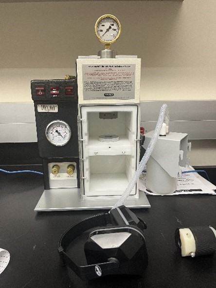
Biolistic particle delivery or micro-projectile bombardment is a technique by which foreign genes are delivered to cells using heavy metal particles coated with exogenous DNA.
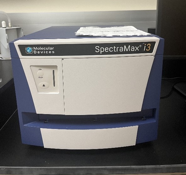
SpectraMax M3 Multi-Mode Microplate Reader is a dual-monochromator, multi-detection microplate reader with full spectral range detection for cuvettes and 6-, 12-, 24-, 48-, 96-, and 384-well microplates.
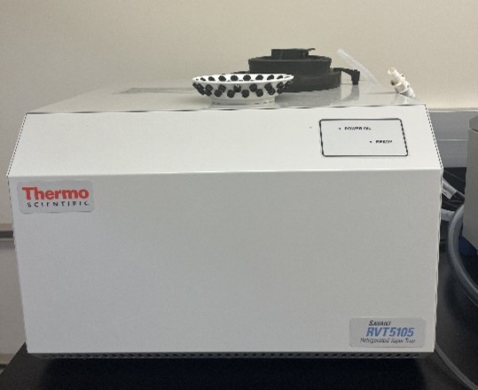
Refrigerated vapor traps for condensation at low and ultra-low temperatures. Solvents are removed from samples during vacuum evaporation.
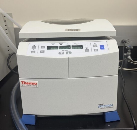
Centrifugal evaporators are used when more than one sample evaporation is required in pharmaceutical, chemical, or biotechnology industries.
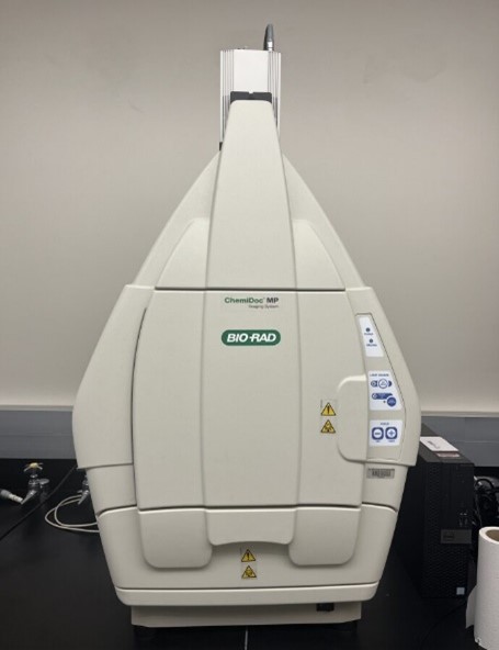
The ChemiDoc MP Imaging System is a full-feature instrument for imaging and analyzing gels and western blots. It is designed to address multiplex fluorescent western blotting, chemiluminescence detection, general gel documentation applications, and stain-free technology imaging needs.
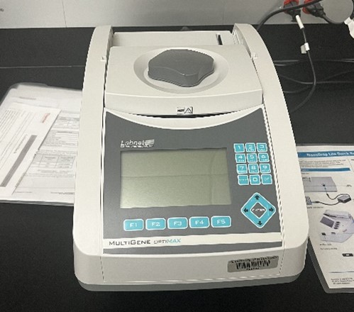
MultiGene Optimax Thermal Cycler is the ideal PCR machine for labs that perform regular genotyping or other high-throughput PCR experiments.
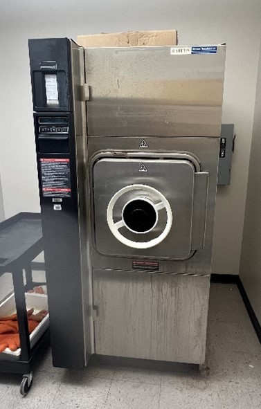
Autoclaves operate at high temperature and pressure in order to kill microorganisms and spores. They are used to decontaminate certain biological waste and sterilize media, instruments and lab ware.
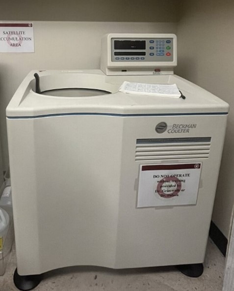
The Beckman J2-HS centrifuge is a refrigerated high-speed system designed for greater throughput and enhanced productivity and can be used for spinning down or samples of variable densities.
The ultracentrifuge is specialized equipment used in laboratories to separate particles, molecules, or cells based on their density. It typically consists of a large circular chamber with a rotor spinning at high speeds to generate centrifugal force.
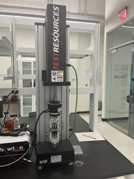
This equipment can be used to perform a wide variety of mechanical tests by pushing in compression or pulling in tension.
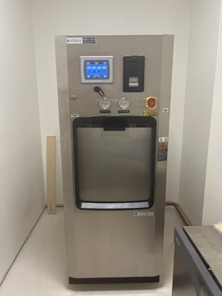
Steam sterilizers are typically used for healthcare or industrial applications. An autoclave is a machine that uses steam under pressure to kill harmful bacteria, viruses, fungi, and spores on items that are placed inside a pressure vessel.
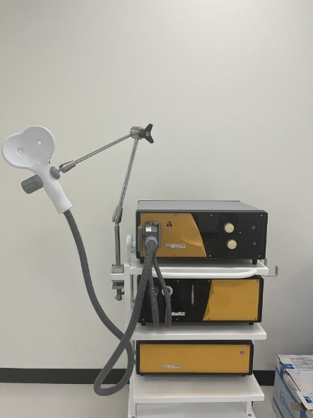
Transcranial magnetic stimulation (TMS) is a procedure that uses magnetic fields to stimulate nerve cells in the brain to improve symptoms of major depression.
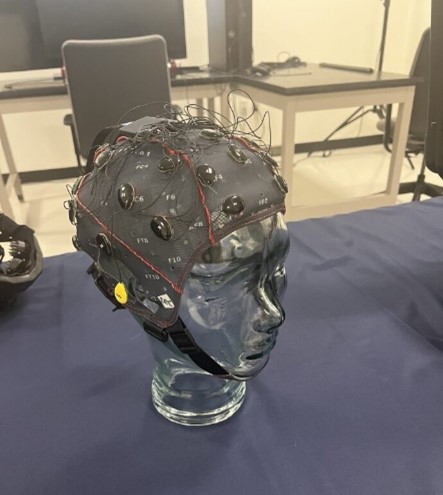
g.GAMMAcap uses active or passive g.LADYbird electrodes and active g.SCARABEO electrodes and has 16 channels. The purpose of the EEG sensors is to monitor electrical activity that exists on the scalp on daily basis due to normal brain activity.
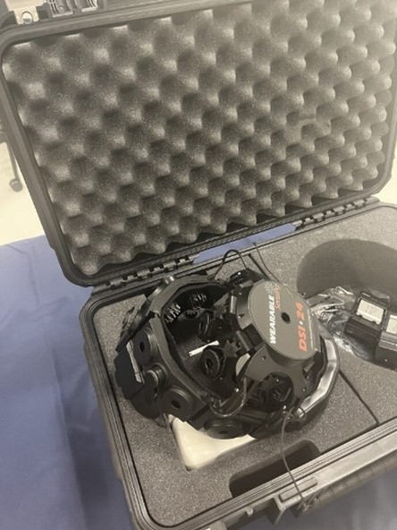
The DSI-24 has sensors that provide full head coverage with 19 electrodes on the head, 2 earclip sensors. Dry sensors allow to get high quality results through hair. That has numerous applications such as biometrics, brain computer interfaces, NeuroEducation, psychological research.
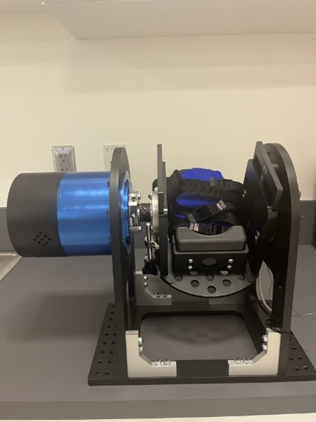
The application of perturbations is externally applied, unanticipated forces used to disturb physical movement patterns. This application has been clinically proven to improve balance, joint stability, postural control and longer-term success with return-to-activity programs.
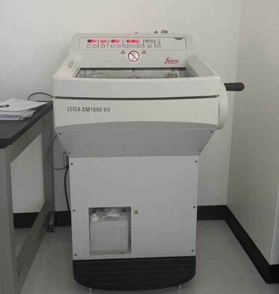
Cryostats are used to preserve frozen tissue samples, slice tissue sections thin enough for microscopic examination, and provide a quick diagnosis for a variety of diseases and medical conditions, including neuromuscular diseases. They can also be used to examine enzyme histochemistry.
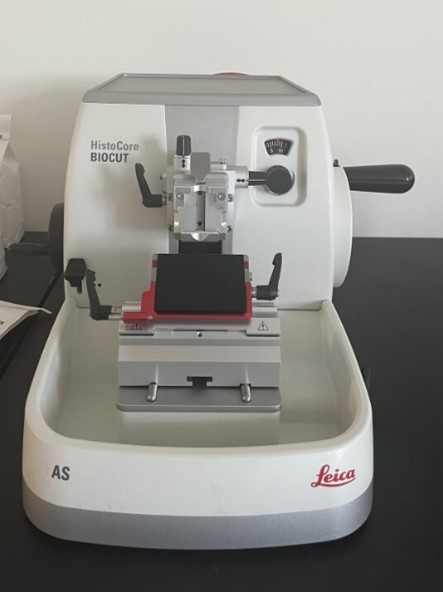
Microtome is a specialized precision cutting instrument, which accurately and repeatedly slices sections from a block of embedded tissue. Different kinds of microtomes are used to section paraffin and plastic embedded tissues.
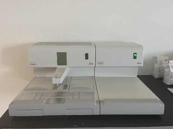
The embedding center is used to surround tissue specimens with paraffin into molds that allow cutting on the microtome. Arcadia C cold plate can be used as a stand-alone unit for re-cooling blocks prior to sectioning or as part of an embedding center to cool mold safter paraffin dispensing.
Microbiological incubators are used for the growth and storage of bacterial cultures.
.jpg)
Thermo Fisher NanoDrop™ One spectrophotometer is both used to perform fast quantification of nucleic acids and proteins.
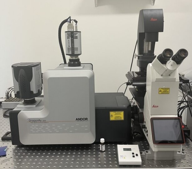
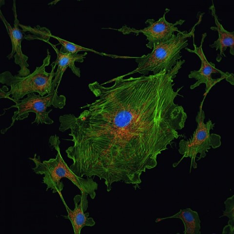
Dragonfly allows researchers to study cancer cell behavior interaction with the environment and spatial distribution of the tumor both in fixed and live cells.
Ultrasound is best used to learn about conditions that involve soft tissues, such as organs, glands, and blood vessels that include small to large animal studies.
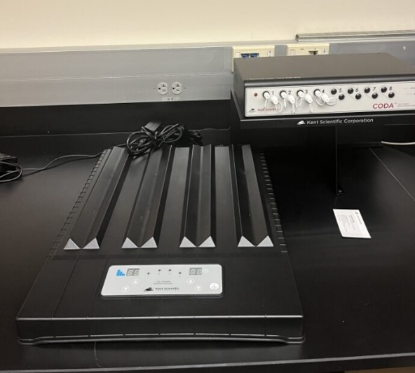
The CODA® mouse rat tail-cuff system was designed to allow accurate blood pressure measurement in mice and rats. Blood pressure is measured in the tail of the mouse or rat using Volume Pressure Recording (VPR) sensor technology.
This center supports light microscopy, transmission electron microscopy, confocal laser scanning microscopy, scanning electron microscopy, focused ion beam microscopy, and X-ray microanalysis of natural and artificial materials. The center provides an array of high resolution microscopic equipment for multidisciplinary and cross-disciplinary science and engineering research and development.
Olin Life Sciences Building, Room 128
Learn more about High Resolution Microscopy and Advanced Imaging Center